Transient Left Atrial Appendage Inversion During Transcatheter Closure Device Placement
Waseem Barham1, Gaurav Dhar 1, Nam S. Cho1, David Rhine1
1Michigan State University College of Human Medicine/Sparrow Thoracic Cardiovascular Institute, Division of Cardiology, Lansing, Michigan .
A 72-year-old female patient underwent left atrial appendage closure. During recapture of the occlusion device, transient inversion of the appendageal wall occurred. We describe the mechanism with real-time imaging and share our experience of handling this situation. To the best of our knowledge, this is the first case report of this unique recapture complication.
Key Words : Left atrial appendage inversion, transcatheter closure, atrial fibrillation.
Correspondence to: Waseem Barham, MD
Division of Cardiology
Michigan State University College of Human Medicine
4650 S. Hagadorn Rd, Suite 100
East Lansing, MI 48823
Left atrial appendage (LAA) closure is a feasible alternative to patients with non-valvular atrial fibrillation at increased risk of ischemic stroke. Although the procedure is generally considered safe, complications still can occur. We report the first case of transient LAA inversion during transcatheter LAA closure device placement.
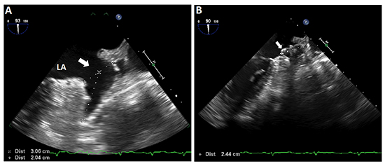
A 72-year-old female with past medical history including persistent atrial fibrillation and essential hypertension was referred for LAA closure. She never underwent any cardiac surgery. She had been on anticoagulation with warfarin for increased risk of cerebrovascular accidents (CHA2DS2-VASc score of 3); However, she had previous massive gastrointestinal bleeding from underlying ulcerative colitis and esophagoduodenoscopy revealed a non-bleeding gastric ulcer. Ultimately, she was deemed a candidate for LAA closure. A standard procedure protocol was followed. Transesophageal echocardiogram (TEE) was used throughout the procedure. Multiplane images and measurements of the LAA were obtained [Figure 1A]. There was no evidence of thrombi or spontaneous echo contrast ("smoke") inside the LAA. Following the preliminary images, right femoral venous access was obtained and transseptal puncture performed. A 14-F delivery sheath (Watchman Double Curve) was then advanced into the left atrium. Through the double curve sheath, a pigtail catheter was then used to engage the LAA and allow for safe advancement of the sheath. After the Watchman closure device was deployed, positioning and compression were verified using TEE and fluoroscopy ([Figure 1B]; Videos 1-3, available online). The position of the device was deemed too proximal to the intended landing zone and compression of the device was less than 10%.
Figure 1. LAA closure using Watchman device. A: Intraprocedural TEE imaging and measurement of the LAA at baseline. There is no evidence of sludge, thrombus or spontaneous contrast. B: The deployed Watchman device (arrow) measuring 4.4 mm in diameter (9.6% compression).
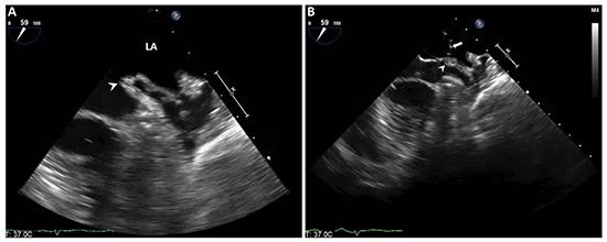
During an attempt at full recapture of the device through the sheath, inversion of the LAA was noticed on TEE and the device failed to retreat into the sheath [Figure 2A]. The distal aspect of the inverted LAA attached to the anchors of the device and appeared as a finger-like heteroechogenic projection pointing towards the endovascular space of the left atrium ([Figure 2B]; Video 4, available online). There was no significant change in the patient's hemodynamic status or evidence of pericardial effusion on TEE.
Figure 2. Intraprocedural TEE imaging during recapture of the deployed Watchman device. A: A finger-like projection (arrow head) captured extending into the LA and represents LAA inversion. B: The Watchman device pedicles (arrow) fixated to the inverted LAA wall (arrow head). LA = left atrium.
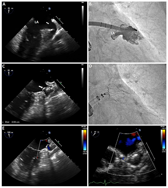
Entrapment of the device and inversion of the appendage resolved after a few series of partial deployment and recapture along with minimal gentle clockwise and counterclockwise rotation ([Figure 3A], Video 5, available online). The retrieved device showed no apparent signs of damage or malfunction. A new Watchman device of the same size was then deployed and released after proper position, anchor, compression and seal were confirmed ([Figure 3B-3E]; Videos 6-8, available online). Postoperatively, anticoagulation with warfarin was resumed. A repeat TEE 6 weeks later showed the Watchman device in place with adequate seal and no peri-device leak. Warfarin was then stopped and aspirin 81 mg daily was started. After 1 year follow up, the patient was doing well and had no symptoms of cerebrovascular accident (CVA).
Figure 3. Closure of the LAA with new Watchman device. A: TEE image of the LAA (arrow) after full device recapture and resolution of the inversion. B: Fluoroscopic appendogram revealing no contrast leakage ruling out perforation of the LAA wall. C: New Watchman device deployed and adequately seated in the LAA with a diameter of 20.3 mm (24.8% compression). D: Fluoroscopic image of the new Watchman device with adequate position and compression. E: Doppler TEE revealed adequate seal with no flow around or across the closure device. F: Six-week follow up TEE shows maintenance of adequate seal and no residual peri-device shunt.
LAA closure has been shown as an effective alternative to anticoagulation to help reduce stroke risk in patients with atrial fibrillation.[1,2] The procedure is generally considered safe and carries low rates of complications including pericardial effusion, air embolism, thrombus formation during device implantation, and device embolization.[3] Left atrial appendage inversion during this procedure has never been reported. Partial or full recapture might be needed to attain appropriate positioning of the device. It is done by advancing the sheath over the deployed device with constant traction. During this process, the LAA luminal surface can potentially remain attached to the anchors and lead to inversion. Inversion of the LAA is generally rare. It has been reported in the majority of the cases following cardiac surgeries, mainly after corrective procedures of congenital cardiac anomalies.[4-9] Rarely, LAA inversion is found without preceding history of cardiac surgery.[10-15] Inverted LAA is often discovered incidentally on postoperative echocardiograms where it appears as left atrial elongated, pyramidal structure and frequently leads to further imaging investigation or surgical exploration.
The unexpected presence often leads to the suspicion of thrombus formation, vegetation growth, tumors, or even foreign bodies. Understanding of the LAA anatomy and comparison with prior echocardiograms can greatly help identify this condition. In our case, the continuous TEE monitoring during recapture highlights a potential hidden complication of recapture, namely atrial appendage inversion secondary to the Watchman pedicle entrapment of endocardial tissue and possible appendage tear with continued sheath advancement.
Operators need to be aware of potential complications during recapturing appendage occlusion devices. Such complications can include appendage tear and acute obstruction of the mitral annular inflow tract with hemodynamic compromise. We emphasize on the importance of careful balance between sheath advancement and traction on the device with continuous monitoring on TEE to detect possible inversion as soon as it develops.
This research did not receive any specific grant from funding agencies in the public, commercial, or not-for-profit sectors.
Inverted LAA may result during LAA closure device position adjustment. The appearance of an intraatrial projection on TEE is very suggestive. Attention is needed to prevent further complications from the inverted LAA.


