Submit Manuscript    >>    Login | Register

Flash News

  •    First placed abstract winner
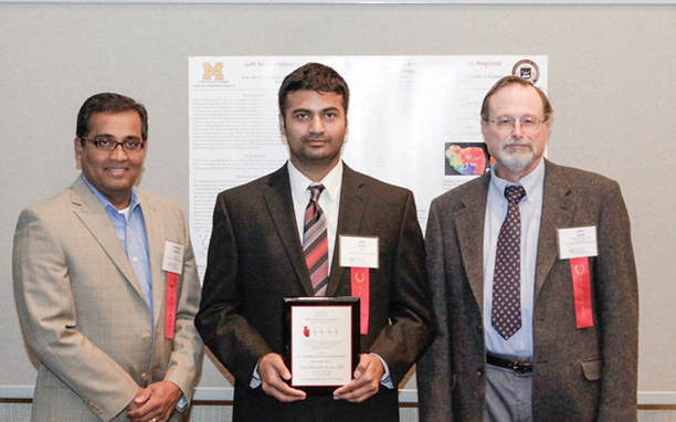
    Dr. Uma Mahesh R. Avula, MD, Research Fellow, Int Med-Cardiology, Center for Arrhythmia Research, University of Michigan.

  • Title : Left Atrial-Pulmonary Vein Infarction leads to Spontaneous Atrial Fibrillation after Regional Action Potential Duration Prolongation
    Authors: Uma Mahesh R. Avula, MD, Masatoshi Yamazaki, MD, PhD, Alvaro Rojas-Pena, MD, Ryan P. O'Connell, BSc, Hassan Musa, PhD, Kuljeet Kaur, PhD, David Demos, M.D, Christopher N. Scipione, MD, Roberto Mondragon, PhD, Raphael Pedro. Martins, MD, Keith E. Cook, PhD, Robert H. Bartlett, M.D, Justus Anumonwo, PhD and Jérôme Kalifa, MD, PhD.
    Category: Cardiac arrhythmias, Basic research
    Introduction: The mechanisms by which atrial infarction results in atrial fibrillation (AF) have not been fully elucidated. Nishida et al. (2011) reported that right atrial infarction is a substrate for AF onset. However, AF mechanisms after left atrial-pulmonary veins myocardial infarction- are unknown. Here, we developed a model of ovine 8-day left atrial-pulmonary veins myocardial infarction (LAPI) and examined changes in local atrial electrophysiology, structural remodeling and AF onset.
    Methods: After thoracotomy, we obtained LAPI in 23 sheep by ligating the left anterior atrial coronary branch. Additionally, 7 sham sheep were obtained without a coronary ligature. Loop recorder was implanted in 17 sheep to monitor for AF episodes. After 8 days, explanted hearts were utilized for: optical mapping, patch clamp, imaging of caffeine-inducible release of intracellular calcium and fibrosis distribution studies
    Results: Ischemic animals presented with more premature atrial contractions, spontaneous AF episodes, and sinus pauses than sham-operated animals. LAPI animals showed increased number of AF episodes that lasted longer than sham animals (p<0.01 for duration between 10 to 60 mins, N=12 LAPI, 5 Sham). Cells dissociated from the ischemic /border zone (IZ/BZ region) showed a significant action potential duration (APD) prolongation compared with non-ischemic zone( NIZ): APD- 98.17±10.33 vs. 46.63±4.67, 127.9±10.62 vs. 73.50±9.45, 181.1±11.26 vs. 119.9±12.19 ms for APD30, 50, 80 respectively (n=7, p<0.005). Caffeine induced calcium peak amplitude (indicative of SR calcium load) and time to 50% decay of peak amplitude did not significantly differ between IZ/BZ and NIZ cells (p=0.07 and 0.39 respectively, N=3, n=9 IZ/BZ, 12 NIZ). Chromanol sensitive current (IKs) was decreased in IZ/BZ cells vs. NIZ by 85% (0.26 pA/pF vs.1.79 pA/pF at 50mV, n=2). Optical mapping of Langendorff-perfused LAPI hearts (n=2), showed substantial impulse conduction delays at the IZ-BZ region. Readily inducible atrial tachycardia and AF episodes (2/2) were maintained by both rotors and breakthrough waves locating at the IZ/BZ. Histological analysis (n=2) showed a significant increase in interstitial fibrosis IZ and BZ vs. NIZ (48.8±28.0% and 27.39±18.29%, IZ and BZ respectively, p<0.05).
    Conclusion: LAPI leads to regional heterogeneity in protein expression, interstitial fibrosis and APD, all of which contribute to the onset of focal discharges and reentries at the left atrial BZ and result in AF initiation and maintenance. Thus, regional LAPI may play a significant role in eliciting left-sided AF electrical sources in patients with coronary artery disease.
Biosense Webster
event date
Introduction to AFib
Ablation Specialist

View Ablation Specialists