A Foreign Material Image In The Coronary Sinus During Coronary Sinus Angiography
Mustafa Yildiz, MD, PhD, Prof1, Gokhan Kahveci, MD2, Yunus Emiroglu, MD2, Okan Erdogan, MD, Prof3
1Department of Cardiology, Istanbul University Cardiology Institue, Istanbul, Turkey.2Department of Cardiology, Kartal Kosuyolu Heart Training and Research Hospital, Istanbul, Turkey.3Department of Cardiology, Marmara University Medical Faculty, Istanbul, Turkey.
Key Words : Coronary Sinus, Angiography, Foreign Material.
Correspondence to: Mustafa Yildiz, Cardiologist, Internal medicine Specialist and Physiologist Department of Cardiology, Istanbul University Cardiology Institue, Istanbul, Turkey.
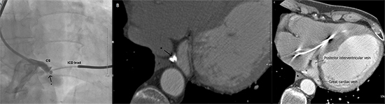
A 63-year-old man with history of bedside temporary pacemaker lead insertion a year ago was hospitalized for cardiac resynchronization and defibrillator device implantation. After insertion of the right ventricular shocking lead we tried to engage the ostium of the coronary sinus (CS) and injected some dye to delineate its anatomy. Unfortunately, the proximal portion of the main CS was occluded. In addition, 2 fixed and rounded neighboring foreign materials were incidentally detected within its opacified portion (Fig. 1A, arrow indicates foreign material). A subsequent multislice computed tomography (Fig. 1B, Fig. 1C* indicates foreign material) confirmed CS occlusion and foreign metallic materials (Hounsfield unit: 2686) resembling metallic electrodes most probably originating from the previous inserted temporary pacemaker lead. We suggested that the forceful blunt insertion of the firm lead tip dissected the CS wall and cretaed a subintimal pouche. Further blood accumulation and formation of intense coagulum externally compressed the wall and occluded the lumen of the CS. When the temporary pacing lead that was partly encapsulated and fixed by the fibrocoagulative tissue was forcefully pulled out, the metallic electrodes overlying the lead tip might have been teared off and retained within the CS. This interesting and rare case highlights an unusual cause of CS occlusion and unexpected complication of temporary pacemaker lead insertion. One must be sure that no fragments have been retained or embolized when removing the pacing leads from the body.1
Figure 1. A foreign material was seen in the coronary sinus angiography (Fig. 1A). A subsequent multislice computed tomography confirmed CS occlusion and foreign metallic materials (Fig. 1B, Fig. 1C; * indicates foreign material)